
Rapid Review: Subdural Hematoma

Sign Up for Free Qbank Access
Get Free Access
Related Articles


Reviewed March 2024
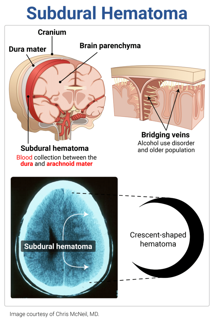
Subdural Hematoma
- Risk factors: traumatic head injury, advancing age, anticoagulant use, coagulopathy, thrombocytopenia
- Caused by tearing of the bridging veins between arachnoid and dura
- Sx: acute or subacute neuro sx, headache, mental status changes, seizures, or focal deficits
- Dx: crescent-shaped hematoma on noncontrast CT
- Management includes neurosurgical consultation, blood pressure control, reversal of anticoagulation
Sample question:
A 68-year-old man presents to the ED after a fall down 12 stairs. On physical exam, you note a large parietal scalp hematoma. His noncontrast computed tomography scan of the head shows a crescent-shaped hematoma. Which of the following is the most likely diagnosis?
Get Free Access and Join Thousands of Happy Learners

You must be logged in to post a comment.
Comments (0)